
Read this blog in:


µTAS 2019 – Miniaturized Systems for Chemistry and Life Sciences
I thought I would write about my experience at µTAS (micro total analysis systems) conference as a newcomer this year. In April, I submitted an abstract of my work and got accepted for a poster presentation.
µTAS 2019 continued a series of conferences that are the premier international forum for reporting the latest research results in microfluidics and lab-on-a-chip technologies, including aspects of microfabrication, nanotechnology, device integration, materials and surfaces, analysis and synthesis, and sensing and detection in the fields of life sciences and chemistry. The conference offers plenary talks as well as contributed oral presentations and posters selected from submitted abstracts. This year it was held in Basel, Switzerland, taking place from Sunday 27th to Thursday 31st October. It gathered more than 1,200 researchers attending and representing more than 400 universities and corporations around the globe. A total of 1139 abstracts and 132 Late News were submitted, 868 of which were accepted including 99 oral presentations. World-renown sponsors, benefactors and exhibitors were also present, including Roche, Dolomite, Ibidi and Novartis, and journals like Lab on a Chip and Analytical Chemistry among many others.
A typical day started around 8-9 am and consisted of plenary lectures, blocks of three parallel sessions to choose from and 2h of poster sessions and exhibit inspections. In addition, we had lunch breaks, industrial stages, workshops, raffles and awards presentations, a PhD student mixer quiz night and a conference banquet dinner.
Final programme and badge.

Photo of the University of Hull team attending the conference.
The poster presentation sessions were meant to be the core of the day, where researchers were able to showcase their work, present them to other peers and get feedback and network with others in a more informal manner. The posters were classified within eight topics that reflected the scope of the growing field, spanning from fundamental physics and fabrication through sensors and detection to applications of microfluidic technology:
- 1. Cells, organisms and Organs on a Chip
- 2. Chemical Applications: Separations, Mixers and Reactions
- 3. Diagnostics, Drug testing & Personalized Medicine
- 4. Fundamentals in Microfluidics and Nanofluidics
- 5. Micro- and Nanoengineering
- 6. Sensors and Detection Technologies
- 7. Other Applications of Microfluidics
- 8. Late News
The work I presented and discussed in the poster session was related to my research on miniaturizing the FISH (fluorescence in situ hybridization) assay on a microfluidic device. FISH is a technique used to detect and identify bacteria and it requires a number of steps using different solutions to treat and prepare the cells and label them with a fluorescent nucleic acid probe that hybridizes within their genome and lights them up. The microfluidic chip used was made of PDMS (polydimethylsiloxane) and consisted of a series of chambers connected by little and shallow gates. The approach relied on a concept known as IFAST (immiscible filtration assisted by surface tension).
In this method, each chamber of the microfluidic chip is alternately filled with the different solutions of the FISH protocol and oil, creating an immiscible barrier on the shallow gates where liquids do not mix. Then, magnetic microparticles coated with antibodies against the bacteria to be detected are used to capture and move them through the gates to the different chambers where each step in the FISH assay takes place. The results I presented were quite preliminary and although they demonstrated a proof of concept for what could be a functional system, a lot of work and optimization has yet to be done. This approach could potentially reduce the time and volumes of reagents used when compared to conventional FISH protocols, facilitate automation and make the assay more portable.
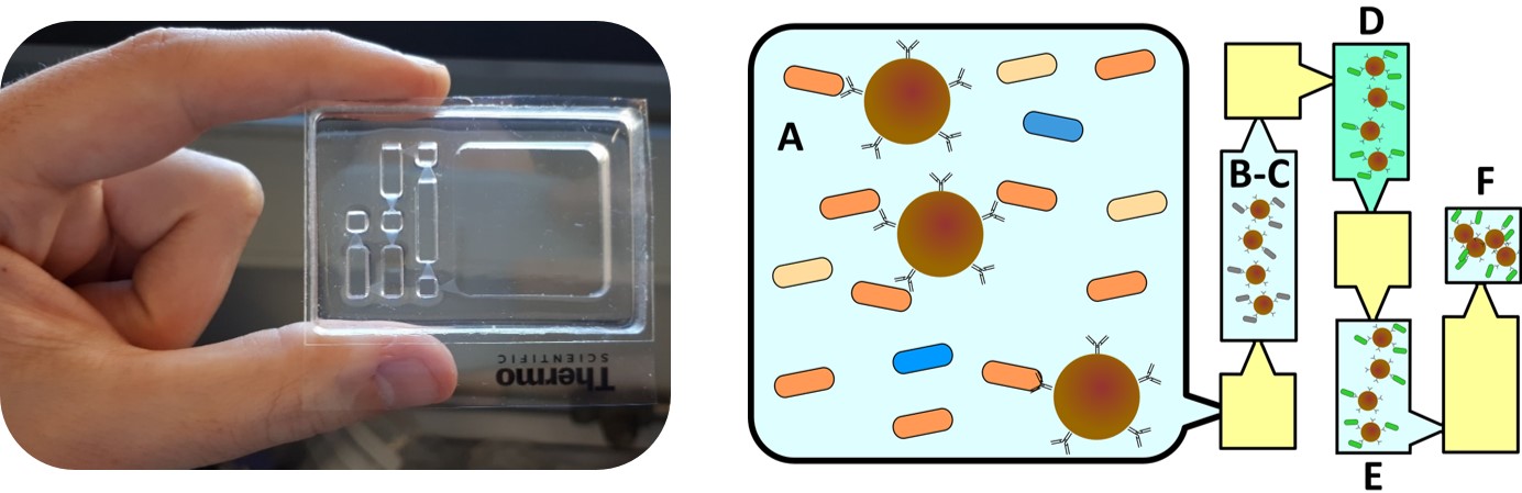
Left: photo of the PDMS microfluidic device used. Right: schematic workflow of the IFAST technique for FISH assay miniaturization. Magnetic beads are used to capture the desired bacteria (A) and move them via external magnet through the different chambers separated by immiscible oil (yellow). Each step of the FISH assay is done in each chamber: fixation/permeabilization (B-C), hybridization (D), washing (E), concentration and collection for microscope readout (F).

Explaining my poster to the jury.
Attending µTAS 2019 with a poster presentation has been a great experience where I have been able to share my research in the leading international conference for microfluidic technologies. I have also done lots of networking and learned about the state of the art in research fields close to mine.
Next year, µTAS 2020 will take place at California, US. I am already looking forward to it!